الخط الساخن 19186
الخط الساخن 19186
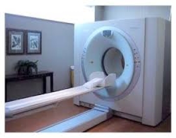
CT or CAT scanning is advanced radiological imaging that uses an x-ray beam that rotates around the patient to capture pictures inside the body. CT images are useful in studies of internal organs because they can separate overlapping structures precisely, producing cross-sectional images of all parts of the body. CT is most used for studies of the head, spine, abdomen, pelvis and chest. The CT scanner also is useful in determining the size and volume of tumors and other masses. This is especially helpful for cancer patients undergoing radiation therapy or surgery.
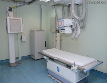
X-rays are a form of electromagnetic radiation, as are radio waves, infrared radiation, visible light, ultraviolet radiation and microwaves. One of the most common and beneficial uses of X-rays is for medical imaging. X-rays are also used in treating cancer and in exploring the cosmos.
Electromagnetic radiation is transmitted in waves or particles at different wavelengths and frequencies. This broad range of wavelengths is known as the electromagnetic spectrum. The EM spectrum is generally divided into seven regions in order of decreasing wavelength and increasing energy and frequency. The common designations are: radio waves, microwaves, infrared (IR), visible light, ultraviolet (UV), X-rays and gamma-rays.
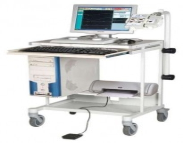
An electromyogram (EMG) is a test that is used to record the electrical activity of muscles. When muscles are active, they produce an electrical current. This current is usually proportional to the level of the muscle activity. An EMG is also referred to as a myogram.
An electroencephalogram (EEG) is a test used to evaluate the electrical activity in the brain. Brain cells communicate with each other through electrical impulses. An EEG can be used to help detect potential problems associated with this activity.
Your doctor may suggest you get an electrocardiogram -- also called an EKG or ECG -- to check for signs of heart disease. It's a test that records the electrical activity of your ticker through small electrode patches that a technician attaches to the skin of your chest, arms, and legs.