الخط الساخن 19186
الخط الساخن 19186
Welcome to Mega Scan radiology centers, we are the first specialized establishment representing the private sector since 1966.
Founded by the pioneer radiology professor Dr. Mohamed Hafez Sherif- the ex president of Egyptian radiology association.
We aim to present & implement the mechanism of experience, accuracy & quality in medical imaging diagnosis by continuous improvement of patient care & to provide up to date technological machines and equipment.
We aspire for internationaly qualified and expert human resource because it is our deep faith that they are truly the ultimate investment to guarantee superior quality services.
We are the first to present radiology centers and ultrasound to Egypt and the MENA region in 1966. Over 50 years ago!
- 1975: first C.T scans in Egypt - Down town and Giza Branch.
- 1995: first open MRI in Egypt.
- 2006: Opening Mohandesin branch with the most up-to-date machines and equipment in medical imaging diagnosis.
- 2016: Opening of Sheikh Zayed branch with the NEWEST WORLD-CLASS international quality medical services
Our slogan: Experience- Accuracy - Quality .
At mega scan works more than 30 doctors, 50 technicians- nurses and admins all together focuses on caring and serving our patients according to principles of professor Dr.Hafez sherif.
- Open MRI.
- PET scanning device (PET / C.T).
- U/S
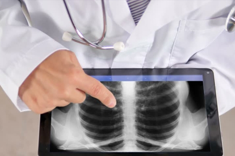
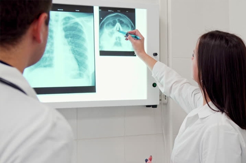
- Xray and normal radiology (all normal rays and contrast).
- Mammography device (early detection of breast cancer).
- Panorama dental.
- ECG and EEG.
- All kinds of laboratory tests in collaboration with Live Lab.
- Echo
- U/S
- Xray and normal radiology (all normal rays and contrast).
- Mammography device (early detection of breast cancer).
- Panorama dental.
- ECG & EEG and EMG.
- Open MRI.
- Multi Slice C.T. Scan.
- U/S
- Xray and normal radiology (all normal rays and contrast).
- Mammography device (early detection of breast cancer).
- Panorama dental.
- EMG.
- Echo.
* Preparation of computerized CT scan on the abdomen and pelvis
1 - abstain from foods that make up the gases the day before the examination such as: vegetables, fruit, soft water, dairy products and eggs.
2 - Allow to eat: pasta, rice, chicken or boiled meat.
3. Gastrografen is taken with 6 cups water as follows:
The initial dose is 12 hours before the test.
The second dose is 6 hours before the test.
The third dose is 3 hours before the test.
4 - Fasting 6 hours before the examination.
5 - Attendance before the date of examination in half an hour.
6 - Bring the previous tests and radiation, if any.
The injected dose in the patient does not pose a risk to the patient's health under normal circumstances. However, there are precautions to be avoided, as in the case of pregnant women, where radioactive material may be a real danger to the fetus, especially in the first months of pregnancy. It should be noted that the patient is the same source of radiation, and this may continue for several hours after the end of the examination (sometimes for 24 hours), in these cases advised not to contact the patient for long periods during those hours, so as not to contact relatives without radiation unnecessarily.